Classically, brain monitoring after SAH can be divided into the time spent on the ICU, with the patient supported by ventilation, and afterwards. Brain monitoring in the ICU does not differ generally from monitoring after TBI. After the ICU period, monitoring is mainly focused on the search of vasospasm and autoregulation of blood flow, mainly using transcranial Doppler ultrasonography (TCD) or near-infrared spectroscopy (NIRS).
As in TBI, ICM+ could be used for high resolution data collection and subsequent analysis of physiological measurement time series in poor grade SAH that undergo neuromonitoring, including ICP, CPP, PbtO2, and cerebral microdialysis.
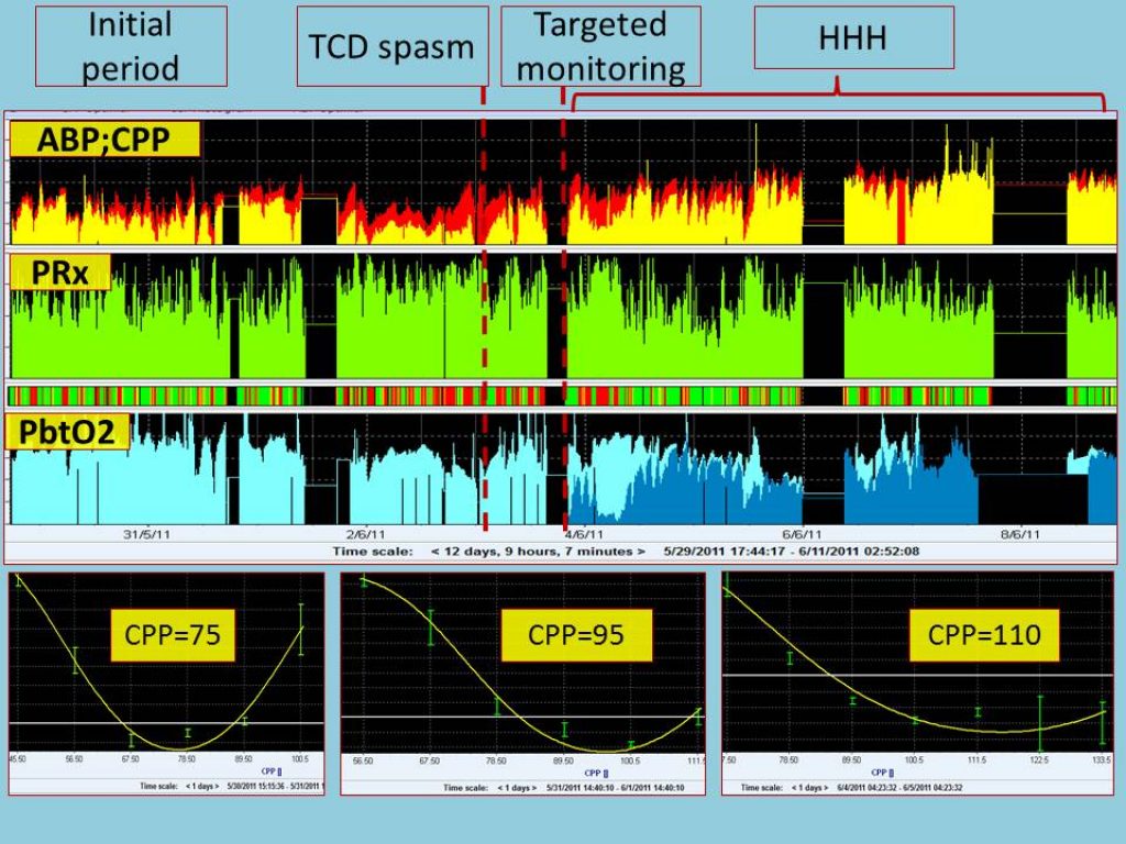
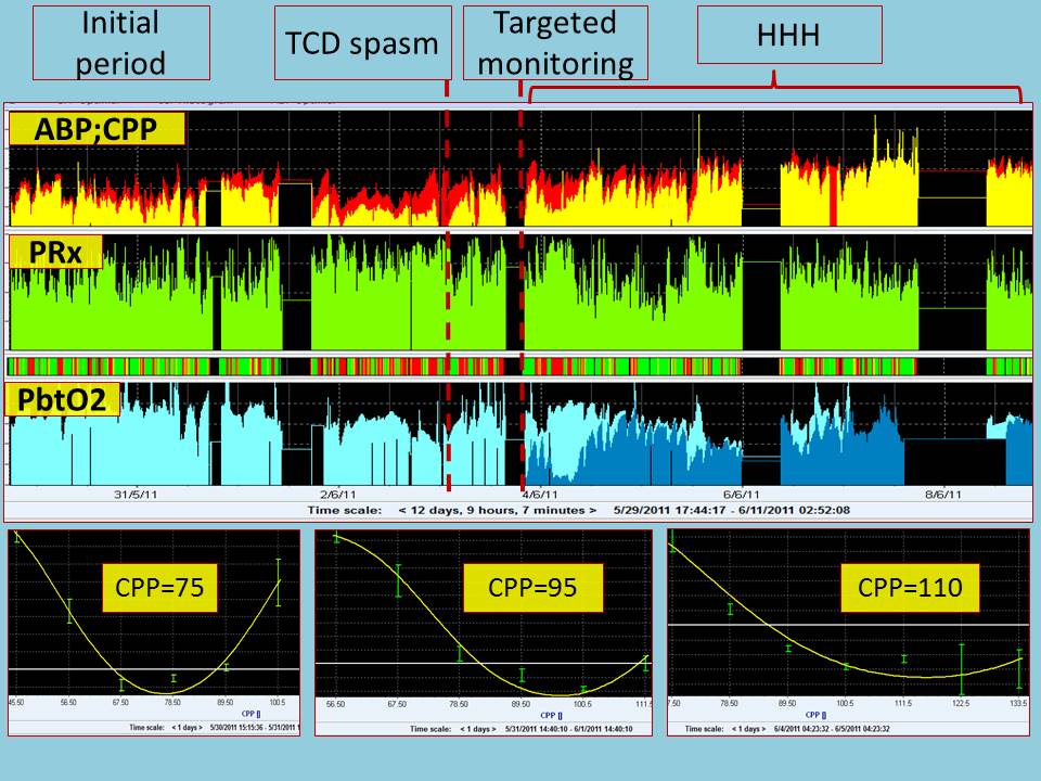
Note that in this case two PbtO2 probes where used, one of them sitting in the affected, spastic, side (dark blue). ‘Optimal CPP’ was calculated and displayed for the initial, spasm, and HHH periods showing gradually increasing value of CPPopt. PbtO2 ipsilateral to the spasm improvement can be observed after introduction of HHH therapy. View image

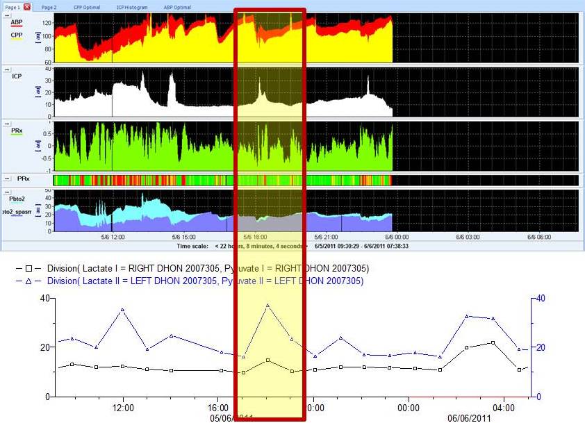
Primary and secondary calculated indices may be compared with cerebral microdialysis recording imported into ICM+. The LPR ratio ipsilateral to the spastic side (blue) and contralateral side (black) show in this example reaction to a temporary decrease in ABP.
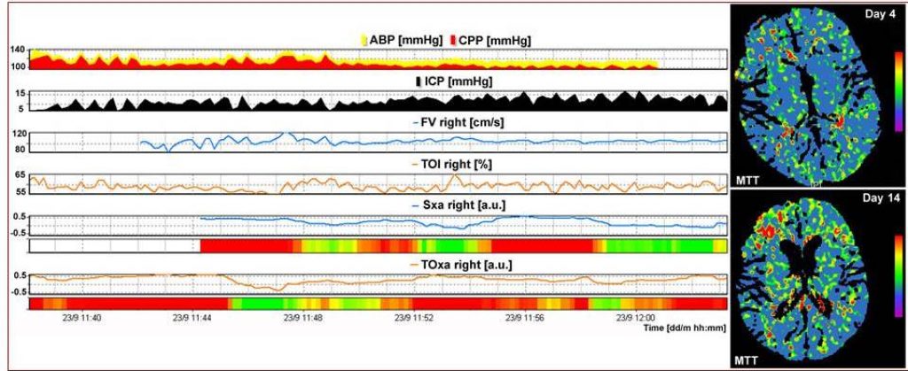
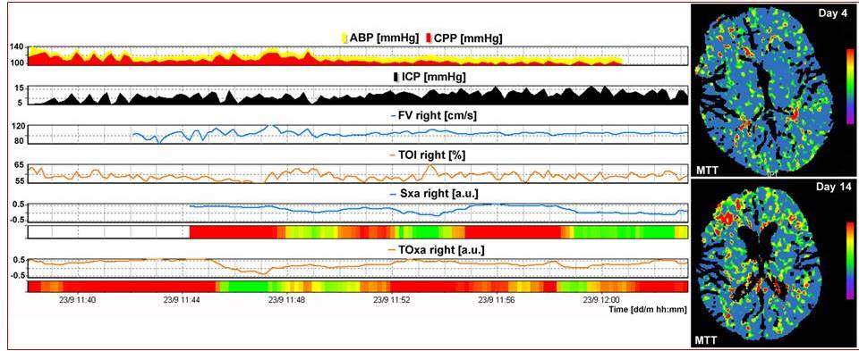
Intermittent monitoring using TCD or NIRS can also be captured and integrated with the other data allowing indices of cerebral autoregulation to be subsequently calculated and analysed.